By Alexis Pasulka, Scripps Institution of Oceanography
February 26, 2010
Preparation for science is officially underway. Today we continued setting up our equipment on the research vessel (R/V) Melville. Now everyone’s finished hustling and bustling around, securing the last of the equipment for our departure at 7:30 tomorrow morning.
We had our first science lecture at 9:00 this morning as part of our at-sea course on reducing ecosystems. All the graduate students and some other scientists gathered in the main lab around our makeshift lecture screen (a white bed sheet secured to the wall with electrical tape). Chris German from Woods Hole Oceanographic Institution talked about the Chile triple junction (CTJ), why we’re heading there, and how our cruise fits into a larger effort to understand reducing ecosystems. As you may have read in our topical essays, the CTJ is a region in which several different types of reducing ecosystems may be found on the seafloor, including hydrothermal vents, cold seeps, large organic falls (whale carcasses), and large organic forms (forests/kelps).

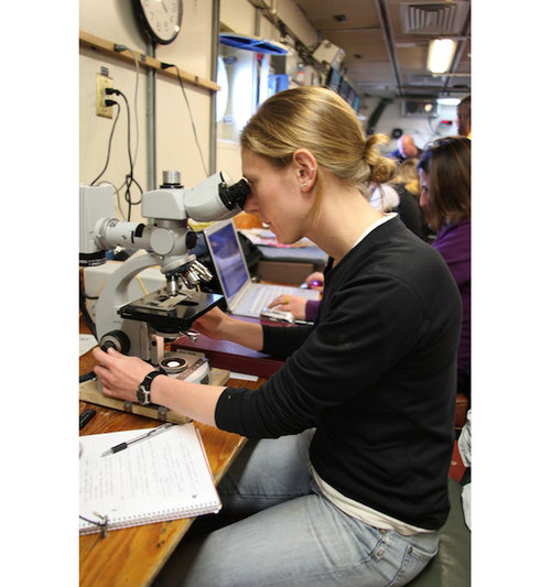
Alexis Pasulka prepares her microscope for the cruise. This involves aligning the halogen bulb and getting the cells on the slide in focus. This microscope has epifluoresecent capabilities, which enables Alexis to look at microorganisms in the water column as well as in the sediments. Image courtesy of INSPIRE: Chile Margin 2010. Download image (jpg, 59 KB).
Following our lecture, we had a safety briefing, two safety drills, and some fun donning survival suits. These suits, which I’m sure are warm and incredibly critical in case of emergency, are quite hilarious to put on. It's even funnier to and probably to watch other people put them on. They’re big red Gumby-looking suits in which you cannot walk or maneuver very well. It made for an exciting afternoon.
In between lectures and safety meetings, I prepared the supplies and equipment for my science. I set up my microscope, finished making chemicals that I will use to stain and fix the microorganisms I collect, and started building an incubator. I’m looking at microbial food-web dynamics at the methane seeps we find, so I will be filtering a lot of water using various filters with different pore sizes.
It’s been a long, but fun day! Tomorrow we should arrive on station and begin our search for plumes.

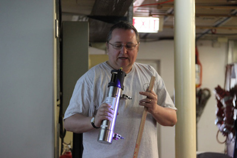
Chris German shows the class a plume miniature autonomous plume recorder (MAPR). This instrument measures depth, temperature, and seawater particle concentrations. In addition, the MAPR has an oxidation reduction probe, which allows us to detect chemically reactive species that would be present in fresh plumes. Image courtesy of INSPIRE: Chile Margin 2010. Download image (jpg, 65 KB).

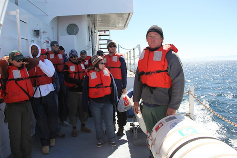
Scientists don life jackets during an abandon ship drill on the upper deck. Ian Lawrence (right), the ship’s first mate, shows the group how to release the life boat in the event of an emergency. Image courtesy of INSPIRE: Chile Margin 2010. Download image (jpg, 91 KB).
I also add various chemicals to the water to stain different parts of the microorganisms. One chemical stains the DNA, while another stains the protein. These stains will allow me to differentiate the organisms under a microscope using a technique called epifluorescence microscopy. By exciting the molecules with a specific wavelength of light, they in turn emit a specific wavelength, which I can see under the microscope. When microorganisms are present, they glow blue, green, red or yellow, depending on the composition of the organisms and which stains were added to the sample.