


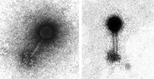
A transmission electron microscope (TEM) image of marine bacteriophage (a virus that infects bacteria). Click image for larger view and image credit.
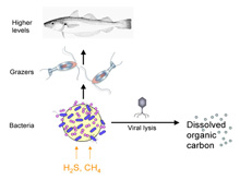
A schematic of the microbial loop/virus loop. Click image for larger view and image credit.
Marine Viruses
July 1, 2007
Chris Kellogg
United States Geological Survey
26° 10.88 N
94° 37.50 W
Most people are surprised to hear that viruses are the most numerous organisms in the ocean. There are approximately 10 million viral particles in a drop of seawater and even higher numbers in the sediment on the sea floor. Nobody panic! The ocean is not a toxic soup of illness waiting to happen! The reason there are so many viruses out there is that there are also a lot of bacteria out there (about 1 million bacteria in a drop of seawater). The majority of marine viruses are bacteriophages, meaning that they only infect bacteria. Some of the most important interactions in the marine environment are taking place at the microscopic level between these bacteria and viruses.
Microbes, specifically bacteria, are recognized as the foundation of deep-sea chemosynthetic communities, both at hydrothermal vents sites and at cold seeps like the ones we are visiting in the Gulf of Mexico. These bacteria are able to use energy obtained from geochemicals (like hydrogen sulfide or methane) at the seeps to generate food from carbon dioxide, in a process that is analogous to photosynthesis in plants. This process is sometimes referred to as the "biogeochemical engine" that drives the chemosynthetic ecosystem. This ability makes bacteria the base of the food web in seep communities. There are symbiotic bacteria providing nutrition directly to the primary macrofauna (e.g., tubeworms, mussels) and free-living bacteria in the waters and sediments around the seeps. Yes, the bacteria are definitely the stars of the show…but the viruses are the directors behind the scenes controlling the story. How’s that? Well, by their ability to infect and kill the bacteria, the viruses control both the bacterial community composition (species diversity) and carbon cycling.
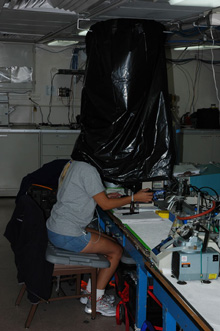
Chris Kellogg isn’t hiding from the rest of the science party; she’s actually using the homemade "darkroom-for-one" to block out the light. Fluorescent dye stains the DNA, enabling her to count virus particles with this microscope. Click image for larger view and image credit.
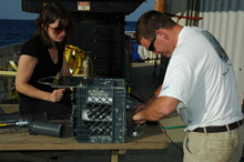
During this cruise, scientists are collecting push-core samples to support a number of different projects related to biogeochemistry, marine viruses, sea-urchin community ecology, and geology. After the samples from the previous dive have been removed and the push cores cleaned and dried, Kate Segarra (left) and Matt Frye prepare nine push cores for the next Jason dive. Click image for larger view and image credit.
For example, if one particular species of bacterium starts out-competing its neighbors and generates a "bloom" (like algae blooms in shallow water), it becomes more likely that a virus capable of infecting that species of bacteria will come in contact with one, starting a cascade of infection. The viruses "crash" the bloom, opening an opportunity for a different bacterial species or combination of species to grow instead. As for carbon cycling in the food web, when a virus kills a bacterium, it bursts the bacterial cell, converting the cellular components into dissolved organic matter (DOM). This transfers nutrients away from grazers and higher levels of a food web, while stimulating other heterotrophic bacterial growth. In this way, viruses can "short-circuit" the food web so that carbon stays at the microbial level instead of moving to larger macroscopic organisms.
Despite that general knowledge, researchers have done little work on viruses in deep-sea environments and no data exists for viruses in cold-seep communities. That's why, during this cruise, I will be sampling sediment cores from a variety of seep environments (e.g., microbial mats, brine seeps, mud flows) to count the number of viral and bacterial particles present. From these counts, calculations can be made to estimate the interactions between the viruses and bacteria (e.g., how much bacterial mortality is due to viruses) and important baseline data will be acquired for these cold seep environments.
Viral particles are very small, often less than 200 nanometers (nm). For perspective, the diameter of a human hair is about 50,000 nm. In the past, the only way to see and count marine viruses was by using a transmission electron microscope (TEM), an expensive and complicated device that uses electrons instead of light to visualize small objects. However, fluorescent dyes have been developed that emit very bright light and these dyes can be used to stain the DNA inside the viral particles. Even though the viral particles are small, the light emitted is bright enough to make them detectable. (Under a portable fluorescence microscope at 100x magnification, the effect is like counting stars in the night sky.) Obviously, the darker the background, the easier it is to see the tiny pinpricks of light.
This type of work is typically done in a darkroom, but most ships do not have a darkroom. And on this ship, where people are working in the lab 24/7, you can’t even wait for everyone to leave so you can turn out the lights. So in a time-honored microbiological tradition, a darkroom-for-one was constructed by suspending black garbage bags from the ceiling to cover the microscope. This blocks enough of the room’s light to allow me to count the virus particles. It does not block the hysterical laughter and camera flashes from my colleagues.