


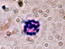
Chromosomes of Lamellibrachia luymesi. Click image for a larger view.
Genome Size of Cold-seep Organisms
October 16, 2002
Peter Deines
Pennsylvania State University
The genetic information that controls the form and function of animals is found in the nucleus of each cell, where it is packaged into chromosomes. We humans have 23 chromosomes in every cell of our bodies. I am a student from Germany, working for a short time in the laboratory of Dr. Fisher at Pennsylvania State University. My aim on this cruise is to examine the number, form and size of the chromosomes of cold-seep organisms, especially the tubeworms. Until now, no one has attempted to karyotype (count the chromosomes) of any siboglinid tubeworm species. We are interested in determining the size of the genome (how much DNA is contained in a nucleus) because this information will compliment other genetic research in our lab. In the end we will try to determine the location of the genes coding for hemoglobin chains on the chromosomes. Hemoglobin is the red substance in blood that carries oxygen. Tubeworms have different kinds of hemoglobins, and it would be interesting to know if the genes for these different kinds are located on the same or different chromosomes. In addition this technique could be also used to compare the rates of cell division in the cold-seep tubeworms and the closely related hydrothermal-vent species. We believe that that worms grow faster in the vent environment; chromosome studies may be used to test this assumption.
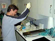
Researcher, Peter Deines is dropping cell suspensions on slides. Click image for larger view.
On the present cruise, we collected tubeworms from several different sites using the submersible. After practicing and modifying the technique of slide preparation with mussels in the lab before the cruise I am now trying to apply this method to tubeworms here at sea. To increase the likelihood of finding dividing cells, young specimens were used in this study. Two different kinds of tissues were tested: gill and vestimentum tissue. After the first preparations, the gill tissue showed much better results. The final chromosome spreading on the slides is the hardest part of this method. Small amounts of cell suspension have to be dropped from a height of at least 30cm onto a slide. On a moving and bouncing ship, this is not an easy task. But after some practice it worked well and we were able to produce the first pictures of a tubeworm karyotype. Back at the lab at Penn State University, high resolution pictures will be taken from the spread chromosomes and then enlarged to determine the exact number of chromosomes.
Sign up for the Ocean Explorer E-mail Update List.