


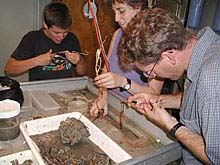
Scientists removing tubeworms from their stiff tubes. Once the worms are out of their tubes, we can collect their eggs and sperm. We then mix eggs and sperm together and they fuse to form embryos. Click image for a larger view.
Development of Cold Seep Tubeworms
Dr. Bruno Pernet
The first submersible dive of the cruise returned to the surface carrying a clump of about 30 large tubeworms. Within three hours a crew of scissor-wielding biologists clustered around a lab table had removed the worms from their tough, woody tubes, laying them out on the table to be measured. Chief scientist Dr. Craig Young then showed us how to “milk” eggs and sperm from mature worms, after which we combined them in dishes of cold seawater to fertilize eggs and start the process of development.
By midafternoon of the first day at sea, we had taken an important first step towards accomplishing one of the main objectives of this cruise—studying the embryology and larval development of deep-sea animals, especially tubeworms. The basics of tubeworm embryology had already been described by Dr. Young and his colleagues, and had helped resolve some long-standing questions about the evolutionary relationships of these peculiar, gutless animals. They found that cold seep tubeworms release eggs and sperm into the water, where they fuse and develop into tiny larvae. On this cruise we are trying to learn more about the biology of these larvae, for several reasons. First, larvae probably spend several weeks drifting in the ocean before they move to the bottom and metamorphose into juveniles. We’re interested in how far they can travel during this planktonic period, and how larvae choose where exactly they are going to settle. Second, newly spawned eggs and sperm don’t seem to contain bacterial symbionts, but adult tubeworms all contain billions of bacterial symbionts that convert sulfide into energy that the tubeworms can use. We want to know how and when larval or juvenile tubeworms manage to acquire these symbionts. Finally, both of the developmental biologists on board—Dr. Young and myself—also just love looking at larvae, and can’t wait to get a good look at these.
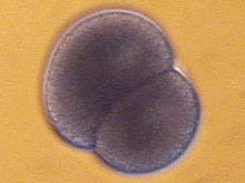
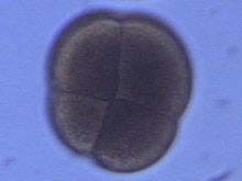
Two-cell (above) and four-cell (below) embryos of the tubeworm Lamellibrachia luymesi. These embryos were fertilized on the first full day of the cruise (7 Oct) and reached these stages about 8 and 12 hours after fertilization, respectively. After about two more days of development, these embryos will form swimming larvae, respectively. After about two more days of development, these embryos will form swimming larvae. Click image for larger view.
The work is conceptually straightforward. We just need to let the embryos and larvae develop to the appropriate stages, look at them alive with a light microscope on board ship, and preserve samples for other sorts of microscopy back on land. In the weeks following the cruise, we will look at developmental stages with a scanning electron microscope (which uses a beam of electrons to “illuminate” the external surfaces of tiny objects) and a confocal laser microscope (which can provide remarkably detailed views of internal structures). In practice, though, studying tubeworm development is sometimes a little tricky. One problem is that it is difficult to get large numbers of embryos, because many develop abnormally. Another is that tubeworm eggs are so rich in lipids (which embryos and larvae use as energy during development) that they float, which complicates standard procedures for handling them. We’re optimistic, though. Cultures of embryos we started the first day of the cruise are still developing, and we’ve been able to start several others since then.
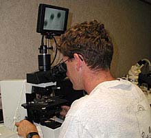
Dr. Bruno Pernet examining tubeworm embryos using a light microscope. The screen at the top of the microscope shows three embryos similar to those shown in the photo above. Click image for a larger view.
Because much of our time is spent waiting for tubeworm embryos to develop, we have time to look at the development of other species that submersible divers collect as well. So far, we’ve made observations of the embryonic and larval stages of two species of snails, and we are trying to obtain embryos of flatworms and “iceworms,” strange little worms that are often found on pieces of methane hydrate. Last night, while most of us were sleeping, the giant fileshell clams, Acesta (see October 8 log) spawned. We are now rearing the babies of that species as well. And we’ve only been at sea for four days!
Sign up for the Ocean Explorer E-mail Update List.