


A typical confocal system consists of a high quality fluorescent microscope, lasers, a control unit, photomultiplier tubes and mirrors, as well as a high-speed computer. Click image for larger view.
Confocal Microscopy
Tara Pitts
Division of Biomedical Marine Research
Harbor Branch Oceanographic Institution
Confocal microscopy is a technique by which digital images of microscopic samples can be constructed. This form of microscopy is typically used for examining samples that can be stained with fluorescent dyes as well as samples that autofluoresce, like the photosynthetic pigments in algae. It can also be used to examine specimens with transmitted light.
A typical confocal system consists of a high quality fluorescent microscope, lasers, a control unit, photomultiplier tubes and mirrors, as well as a high-speed computer. The system requires a cool environment, a vibration-free platform, and an area that can be darkened. The confocal operator must have basic knowledge and skills in microscopy. The operator must also be trained on how to use the system as well as the specialized software.
The confocal system focuses on one plane of view at a time and records a digital image. All the planes are combined to form a focused three-dimensional image. Special software allows for viewing the entire image or each focused plane. The advantage of using confocal microscopy over standard fluorescent microscopy is this feature that enables the scientist to examine a cell or structure in 3-D. The older technology only allows for viewing one plane of view at a time, while the rest of the image is often out of focus. Confocal microscopy allows one to see the whole specimen or parts of the whole specimen in a sharp 3-D image. After acquiring this digital image the software allows manipulation of the image for better intensity or to adjust the quality of the image. This digital image can be saved as a file that can be imported or exported for use in other programs. It produces quality images that can be used for publications, posters, presentations or even web sites.
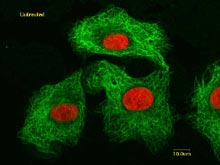
Untreated cancer cells. Microtubules which form the cytoskeleton are stained green (with a fluorescent stain called FITC). The nuclei are stained red (with a fluorescent stained, propidium iodide, which is specific for nucleic acids). Fluorescent image. Click image for larger view.
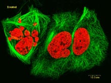
Drug-treated cancer cells. These cancer cells were exposed to a drug for 24 hours before they were fixed and stained for viewing. Cytoskeleton is stained green; nuclei are stained red. Click image for larger view.
Using confocal microscopy, scientists can study both living and preserved samples. One application for which confocal microscopy has been particularly useful is evaluating the effects of chemicals produced by marine organisms on specific structures or processes in cancer cells. This has helped scientists determine the mechanism by which these chemicals kill cancer cells, and could lead to the development of new drugs to treat cancer. Another application of this technology is the identification of unique microorganisms that live in marine invertebrates (such as sponges).
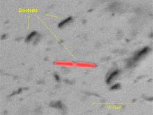
Bacteria from a sponge cell suspension showing some autofluorescent bacteria. Transmitted light and Krypton laser. Click image for larger view.
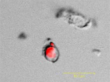
Autofluorescent microorganism from a sponge cell suspension. Transmitted light and Krypton laser. Click image for larger view.
In some cases, the bioactive chemicals may be produced by microbial symbionts, and confocal microscopy could be useful in identifying the type and location of the microbe in its host.