


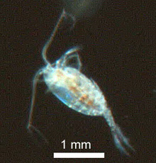


Fig. 1. Digital holographic imaging apparatus in the ship's lab. Image courtesy of 2007: Exploring the Inner Space of the Celebes Sea.
Digital Holographic Imaging
October 6, 2007 — Log 1
Nick Loomis
Graduate Student, MIT
3D Optical Systems Group
Russ Hopcroft has kept us busy with plankton tows ever since we left Manila. Many interesting zooplankton come to the surface at night to feed in safety, which means that my bedtime has gotten progressively later and later. Thank goodness for the creatively strong coffee brewing in the mess hall!
Sampling plankton with nets has several drawbacks. The numbers of delicate objects — such as "marine snow," Trichodesmium, and gelatinous animals — are vastly underestimated. It's also impossible to pinpoint exactly where different species are located, which may vary within just a few meters. Another big challenge is that someone has to sit down, categorize, and count everything collected. Cabell Davis' video plankton recorder (VPR) is a device for collecting information on undisturbed plankton living in the ocean. My group at MIT has been developing another technology, digital holographic imaging (DHI), to study undisturbed marine plankton.
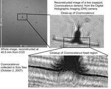
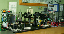
Fig. 2. An image of the copepod Cosmocalanus dawinii, reconstructed with the digital holographic imaging (DHI) system from the in situ recording made by the video plankton recorder. In the close-up shot, feathery feeding appendages are visible on each side of the head, as are the chemical sensing and tactile hairs on the antennae.
DHI is a special form of three-dimensional (3D) imaging. We use a controlled laser to illuminate a plankton sample, then record the light which scatters off the plankton onto a 16 megapixel digital camera chip (fig. 1). Using our knowledge of how light propagates, we can then work backward to calculate the "objects" which created the scattering pattern. Using our algorithms, we can look at any slice in the sample volume with 9 micron resolution — it's like having a microscope that can be focused through a huge volume at any time after the hologram is recorded, so there's never any part that's out of focus. Our laser is active only for a few microseconds at a time, so fast-moving appendages — cilia and podae — are "frozen" in time (fig. 2).
Our first goal is to record digital holograms in the lab from samples collected with our nets and with the remotely operated vehicle (ROV). The holograms include information about small things that get overlooked by the microscopists, along with "larger" animals such as copepods (hundreds of microns to a few millimeters in length). We're also comparing our hologram images with images that Russ records though his video microscope and Cabell with his video plankton recorder. Our final goal is to test a prototype low-power DHI system. We actually recorded our first good copepod image with the prototype last night, which means we're on track to put the DHI system into an underwater housing as soon as we return home.
From the perspective of an optics guy, this cruise has been a great chance to learn about ocean biology from the experts. It's also been a great view into how other optical tools are used to image marine samples, including video microscopes, ROV and RopeCams, and photos of live plankton in specialized lab aquariums called kreisels.