Coral Ecosystem Connectivity 2014

Mission Plan

By Robert Cowen, Ph.D., Director and Project Lead - Hatfield Marine Science Center, Oregon State University

R/V F.G. Walton Smith: August 14-28, 2014
M/V Spree: June 18-27, 2014
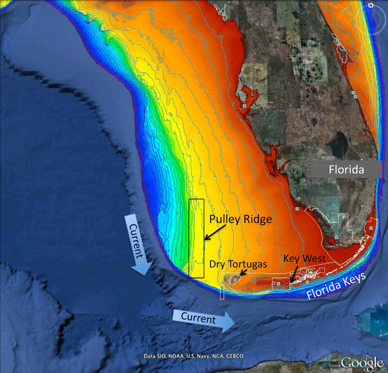
Figure 1. Map of project area showing Pulley Ridge, off the west coast of Florida at depths of 200-330 feet in relation to the downstream refs of the Dry Tortugas and Florida Keys. Colors represent water depth, which ranges from 33 feet (red) to depths of 820 feet or greater (dark blue). Current arrows depict prevalent current direction. Background image is from Google Earth and the depth information is from the U.S. Geological Survey and NOAA.

Figure 1. Map of project area showing Pulley Ridge, off the west coast of Florida at depths of 200-330 feet in relation to the downstream refs of the Dry Tortugas and Florida Keys. Colors represent water depth, which ranges from 33 feet (red) to depths of 820 feet or greater (dark blue). Current arrows depict prevalent current direction. Background image is from Google Earth and the depth information is from the U.S. Geological Survey and NOAA. Image courtesy of Robert Cowen. Download larger version (jpg, 1.4 MB).

This is the third year of a five-year study to investigate the role that the mesophotic reefs of Pulley Ridge (off the southwest coast of Florida) may play in replenishing key fish species, such as grouper and snapper, and other organisms in the downstream reefs of the Florida Keys and Dry Tortugas (Figure 1).

Mesophotic reefs are coral reef environments found at depths ranging from 30-40 meters to greater than 100 meters in the Gulf of Mexico where sufficient light enables certain reef-building corals (i.e., corals with symbiotic algae growing in them) to survive. Mesophotic reefs support a diversity of populations of algae, sponges, corals, other invertebrates, and fishes.
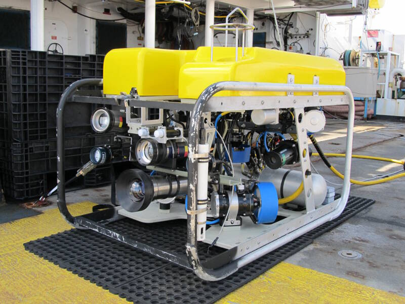
Figure 2. The R/V F.G. Walton Smith, owned and operated by the University of Miami, Rosenstiel School of Marine and Atmospheric Sciences (RSMAS), is one of two vessels that we will be using during this expedition.

Figure 2. In 2014, we will be using a new remotely operated vehicle, the SubAtlantic Mohawk 18, owned by the National Marine Sanctuary Foundation and the Flower Garden Banks and operated by the Undersea Vehicles Program at the University of North Carolina Wilmington (UVP/UNCW). Image courtesy of UVP/UNCW. Download larger version (jpg, 1.9 MB).

In the summer of 2014, we are conducting our third year of fieldwork, using two separate vessels. The first cruise took place on the M/V Spree (a charter dive vessel operated out of Key West, FL) from June 18-27; read a summary here. The second cruise will occur on the R/V F.G. Walton Smith (owned and operated by the University of Miami/RSMAS) from August 14-28 (Figure 2). A total of seven science divers and one technician were on the Spree, while 12 scientists and technicians will be on the Walton Smith. Each vessel will address a different facet of the project.

M/V Spree Cruise

On the Spree, the science divers focused on collecting specimens of target taxa for population genetic analyses in the laboratory. We made these collections in areas that have been shown to support higher-than-average densities of the target species.

All specimens will be used for genetic studies and age and growth information for the fish species. Additionally, the divers recovered and redeployed instruments that track oceanographic currents during this mission.

R/V F.G. Walton Smith

On the Walton Smith, the science team will focus on:
  • Characterizing the benthic (bottom) and fish communities using a surface-driven remotely operated vehicle (ROV). A high-definition still frame camera mounted on the ROV and pointed downwards will record the abundance and distribution of benthic organisms and bottom type (e.g., sand, broken coral rubble, and live bottom). A video camera pointed forward will record fish species present and their abundance in front of the ROV as it moves along the bottom. By simultaneously recording the GPS position of the ROV, we can estimate the area of the bottom actually covered and equate the abundance of the various organisms to density estimates.
  • Collecting genetic samples of larger fish species using fish traps. Some fish are too large and fast or too rare to adequately capture via the ROV cameras – this is especially true for the deeper groupers. These fish are more easily sampled using fish traps. Traps will be lowered to the bottom during the early evening and remain on the bottom throughout the night. At dawn, the traps are brought back up to shallower water where a diver will inspect each trap and if any grouper are present, the diver will deflate the fish’s swim bladder (to prevent damage to internal organs from changing pressure), then take length measurements and sample a small portion of the caudal fin for genetic studies.
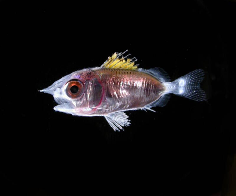
Figure 3. A larval squirrelfish of the Family Holocentridae. Larval fish are sampled using plankton nets or light traps.

Figure 3. A larval squirrelfish of the Family Holocentridae. Larval fish are sampled using plankton nets or light traps. Image courtesy of Cedric Guigand. Download larger version (jpg, 359 KB).

  • Characterize planktonic larval fish and invertebrates using an imaging system and light traps. As we are interested in the dispersal potential of these various populations, we must pay particular attention to the larval (young) stages (Figure 3), since this stage is often where dispersal occurs. This is especially true for species that remain closely affiliated with (or attached to) the bottom as adults. To achieve this, we sample the plankton (where the larval stages reside) using an imaging system and light traps – each method serves to capture information about different species. For example, we use an imaging system (known as the in situ ichthyoplankton imaging system, or ISIIS) to look at fine-scale distribution of larval fish (and their prey) from the surface to near the bottom and we use light traps to capture the larval fish as they end their larval stage and ‘settle’ to the bottom as juvenile fish. These specimens are also used for genetic studies and additional biological analyses (e.g., age and growth) back in the laboratory.