


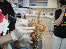
Manning Seamount marine life, found on an experimental basalt block and collected using the Hercules bio box. ![]() Click image to view a slide show.
Click image to view a slide show.
The Caprellid Dance
May 15, 2004
Mary Grady
Adjunct Earth Science Instructor
Northeastern University
Diana Payne
Biologist and Education Coordinator
Connecticut Sea Grant
When you do dance, I wish you
A wave o’ the sea, that you might ever do
Nothing but that.
—William Shakespeare, The Winter’s Tale, Act IV, Scene 4
Today’s first remotely operated vehicle (ROV) dive was shorter than previous dives, starting around 9 Friday evening and ending near 8:30 this morning. Although additional dives are planned on the Manning Seamounts, the science and ROV team decided that it was best to ascend and move the ship to the next dive site, instead of driving the ROV there.
Through late afternoon yesterday, Institute for Exploration (IFE) team members Dave Wright and Webb Pinner designed a new claw for the manipulator arm, "Predator," to make the sampling process more effective. Dr. Peter Auster, watch leader for the midnight to 4 am shift, noted that the new claw worked well. “It was great,” said Dr. Auster. “It was built to collect coral. It saved a lot of time, and the samples were collected in better condition.”
The Manning Seamounts are located at 38º N latitude, 60 W longitude. At more than 90-million-years old, they are among the youngest seamounts we will visit on this cruise. Actually, Manning is a set of five, closely clustered seamounts. The minimum depth to reach the shallowest seamount summit is 1,504 m. The actual height of the tallest Manning seamount, as it rises from the abyssal plain, is approximately 3,500 m
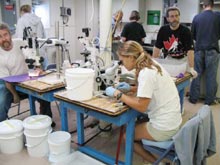
Les Watling, Katie Olds, and Scott France work to process samples. Click image for larger view.
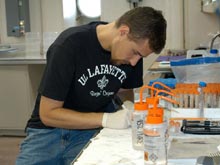
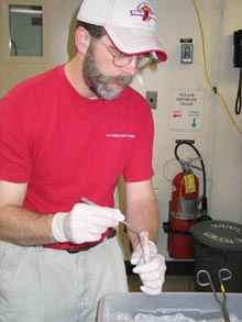
Mercer Brugler prepares coral specimens. Click image for larger view.
In a laboratory aboard the Ronald H. Brown, many of the scientists are crammed into a relatively small space. Dr. Scott France and graduate student Mercer Brugler prepare coral specimens for DNA analysis. Graduate student Anne Simpson prepares her coral samples for analysis of the reproductive structures. Dr. Lauren Mullineaux and research assistant Susan Mills examine samples to learn more about organism dispersal and colonization. Susan is also examining and photographing several interesting worm specimens.
The team members remove coral specimens from the cold room one bucket at a time. Each specimen is brought into the lab for the science team to photograph and take samples. Paragorgia was fairly abundant (in a variety of colors) on today’s dive.
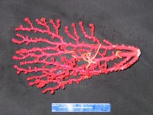
Red Paragorgia coral collected from one of the Manning Seamounts. Click image for larger view.
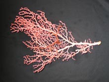
Pink Paragorgia collected from one of the Manning Seamounts. Click image for larger view.
In the lab, scientists took turns looking at an unusual coral specimen under the microscope. “That’s weird!” said one after another, as they studied the unusual morphology. “Instead of the normal polyp shape of a mouth surrounded by six tentacles, these are kind of squashed along the branch,” Dr. France said. “And there is some kind of tube sticking out of their mouths.” Mercer guessed the tube was some kind of worm, but others were skeptical. Dr. Watling was called over for his opinion. His first reaction was the same as everyone's: “That’s weird!” He judged that since every one of the polyps had a tube in its mouth, the worm hypothesis wouldn’t stand up. Close-up video studies were the next step in gathering more information. Initial studies suggested that the "worm" was actually digestive mesenteries, or flaps sticking out of the coral-polyp mouths.
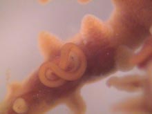
Microscopic image of digestive mesenteries sticking out of the mouth of a coral polyp. Click image for larger view.
Despite the long hours and the unending work—or maybe because of it—spirits are high as the scientists preserve and investigate their samples. Susan Mills projects her microscope onto a large video screen, where everyone can admire the images, showing incredible details of a variety of worms and invertebrates. “That’s a caprellid,” said Dr. Jon Moore. To demonstrate its behavior, he and Mercer invented the “Caprellid dance,” holding up both hands and waving them, while shifting hips from side to side. This made clear to everyone what kind of organism they were looking at. (“Oh yes, the caprellid!”) Later, Mercer adapted the technique to demonstrate worm behavior, while watching some under the microscope. These activities, known as the "late night sillies," are common on cruises where scientists work long hours on things they enjoy!
Dr. Scott France puts a coral tissue sample into fixative for DNA studies. Click image for larger view.
It’s in the Box
Lance Arnold
Tolland High School
Tolland, Conn.
A major objective of the Mountains in the Sea expedition is to study the biodiversity of seamount organisms, particularly corals. Scientists on board are interested in collecting the soft corals, which are found on seamounts thousands of meters below sea level. The corals can be used for a variety of studies.
Dr. Scott France is interested in soft coral genetics and how the corals are related to each other. He collects tissue samples from the corals for DNA analysis to sequence mitochondrial genes.
Dr. Les Watling hopes to understand which corals may be found on different seamounts that are isolated from each other, as well as what organisms are associated with the corals. He samples and preserves collected coral pieces and any organisms found attached to them.
It is important for these scientists to collect healthy, live deep-sea coral specimens and to bring them to the surface, protected in a closed insulated container known as a bio box. The soft corals degrade rapidly in water a few degrees warmer than that in which they live. The sequence of events involved in acquiring the specimens for research can be tricky. Getting the corals into the bio box is crucial.
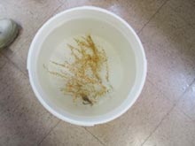
A feather star and brittle star share a bucket of ice-cold water with the coral sample they were attached to. Click image for larger view.
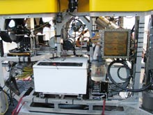
A front view of the ROV Hercules shows the white bio-box, powerful arms, lights, and cameras. Click image for larger view.
Dr. Scott France examines a piece of coral and removes a worm for later analysis. Click image for larger view.
Beyond a ship capable of mid-ocean travel, scientists also need a vehicle—manned or unmanned—to get to the deep-ocean floor. This vehicle must be equipped with tools both to collect the corals and to place them in the bio box. Cameras and lights are also crucial components of these underwater systems. The ROV Hercules, with its suction and manipulator arms, video cameras, and ability to dive thousands of meters fulfills the requirements.
Once the bio box is at the surface, the team members move the organisms to waiting buckets of ice-cold water and then into a cold room. From the cold room, specimens are taken to be photographed against a black background. Samples of corals are studied and pieces are collected into fixatives for DNA studies back on shore. Ideally, organisms are collected from the bottom, photographed, and processed in a matter of hours.
Sign up for the Ocean Explorer E-mail Update List.